
All-in-One Fluorescence Microscope
BZ-X series
All-in-One Fluorescence Microscope BZ-X series
No Darkroom Required
Streamline Imaging and Analysis with a Single Platform
Simple setup and easy operation for outstanding research results. Capture confocal-quality images faster, more easily, and with less damage.
- Built-in Darkroom, Space Saving
- Any User Can Easily Capture Images
- Image an entire well plate for high-throughput analysis
- Referenced in over 15,000 publications
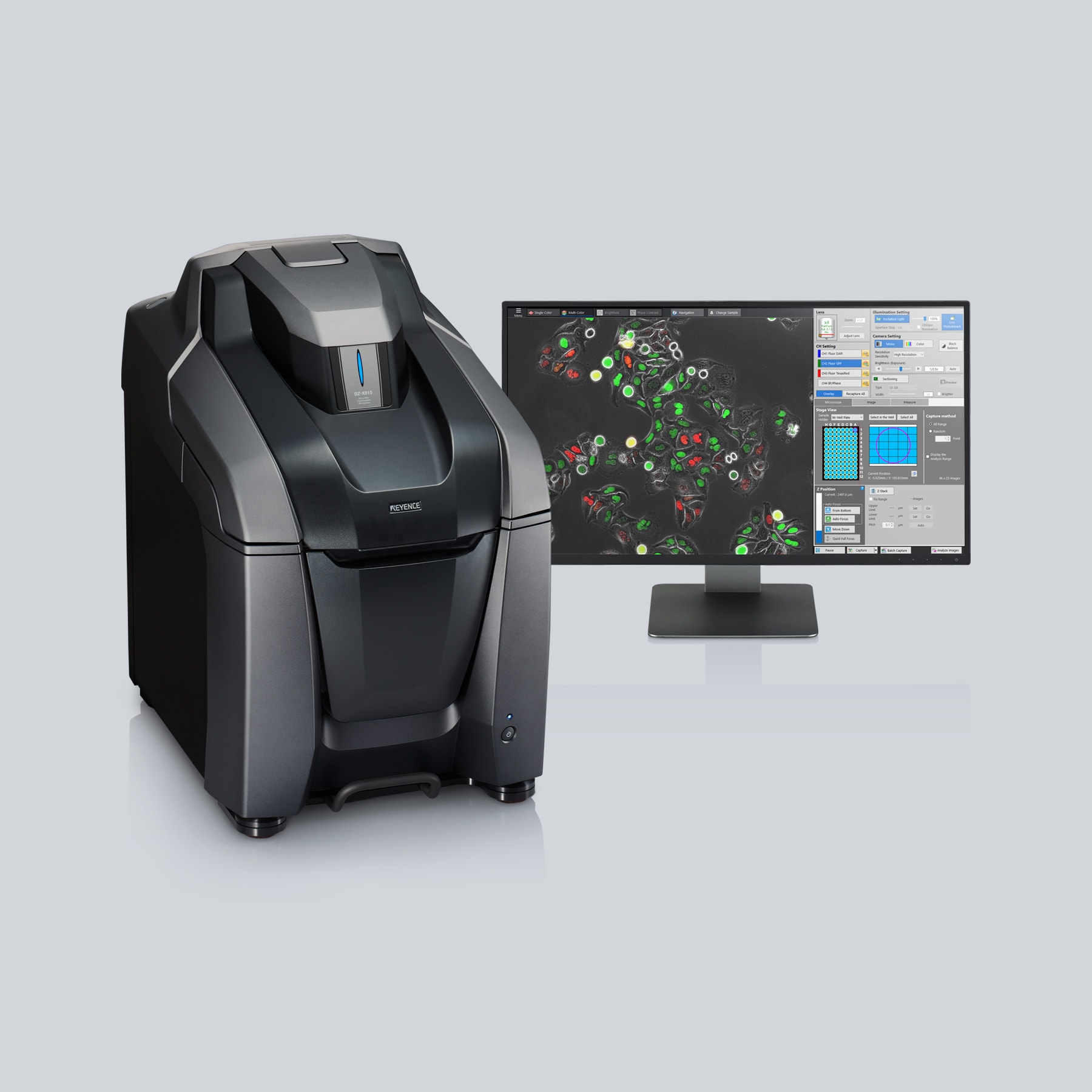
The BZ-X Series is an all-in-one fluorescence microscope equipped with advanced observation capabilities, a fully-motorized control system for instant target acquisition, and advanced imaging and analysis functions. With a built-in darkroom and an installation area of ~ 1' x 2' the BZ-X inverted fluorescence microscope can be set up in any location for optimal testing efficiency. The high-sensitivity, cooled CCD camera can be switched between monochrome and color imaging, and supports fluorescence, brightfield, and phase contrast imaging. The large motorized stage can quickly move to the desired observation location, while high-speed auto-focusing and automatic capture conditions allow any user to easily capture publication-quality images. Additionally, the BZ-X Series all-in-one microscope is capable of three-dimensional analysis, motion analysis, and time-series quantification.

Features
Capture Clear Images Without Fluorescence Blurring
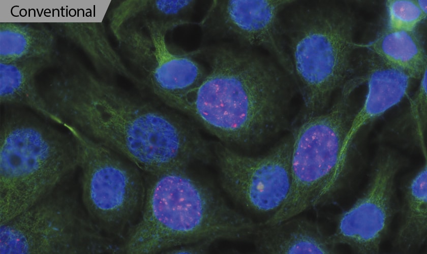
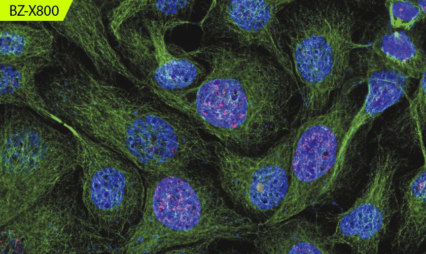
Optical Sectioning
Easily capture high-definition images without the blurring caused by out-of-focus signals with this all-in-one microscope. The unique optical sectioning technology in the BZ-X800E uses an electronic projection element for structured illumination. Operation is simple and intuitive, allowing even first-time users to capture publication-quality images in seconds.
Tubulin and H2AX Courtesy of Momoko Ishikawa, Department of Pediatric Dentistry, Tohoku University Graduate School of Dentistry
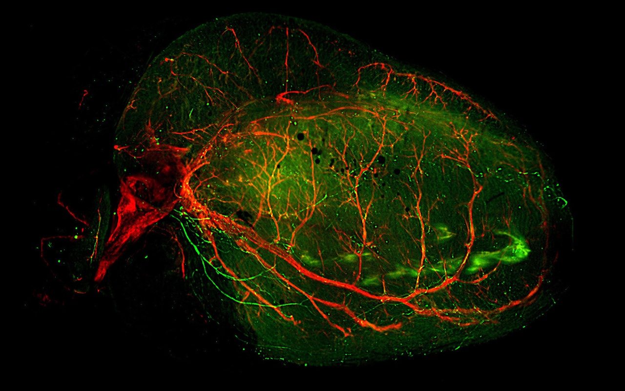
High-Speed Capture of High-Resolution, Wide-Area Images
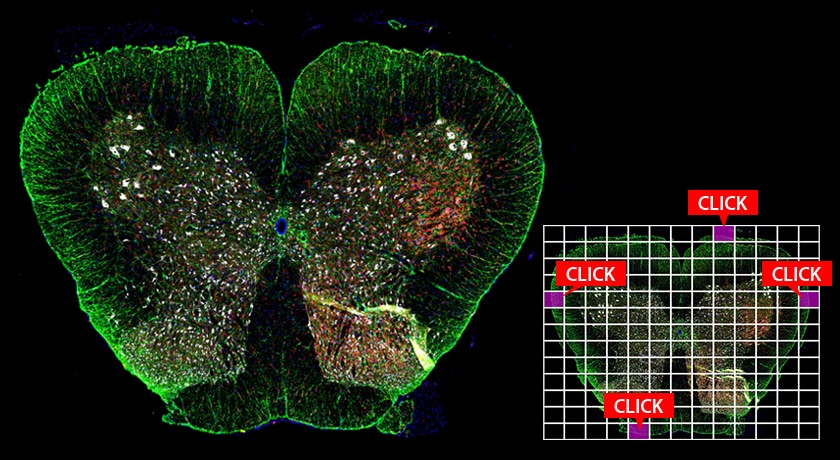
Rat spinal cord
Courtesy of Professor Tasuku Nishihara, Department of Anesthesia and Perioperative Medicine, Ehime University Graduate School of Medicine
Image Stitching
Viewing a specimen at high-magnification often requires an expansion of viewing area beyond a single field of view. Image stitching in this wide-field microscope allows the user to easily capture an entire specimen at high-magnification, and seamlessly create a single high-resolution image.
Up to 50,000 x 50,000 pixels can be rapidly joined together without stitch lines or brightness variations. A large quantity of images can be captured at a speed that is seven times faster than that of conventional methods.
Image Cytometer Module
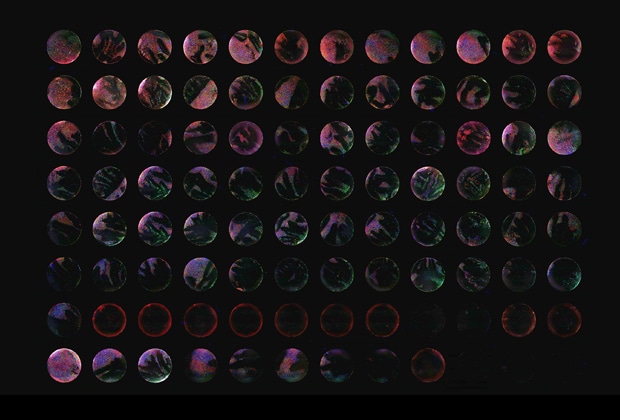
High Throughput for Capture and Analysis
Capture settings in one location can instantly be applied to all fields of view on a well plate. Users can select any or all wells to be scanned with uniform conditions for high reproducibility of data. This work flow can be completed in just three simple steps. The system will then automatically execute the capture without any additional user configuration.
Image Cytometer Analysis
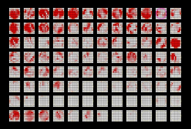
Accurate, High-Content Analysis with High-Resolution Images
Set analysis conditions for a single image and apply to all data points automatically. This saves time and reduces variability from one image to the next. The BZ-X800E's advanced optics capture high-resolution images, resulting in highly precise data acquisition.
BZ Series Application Examples
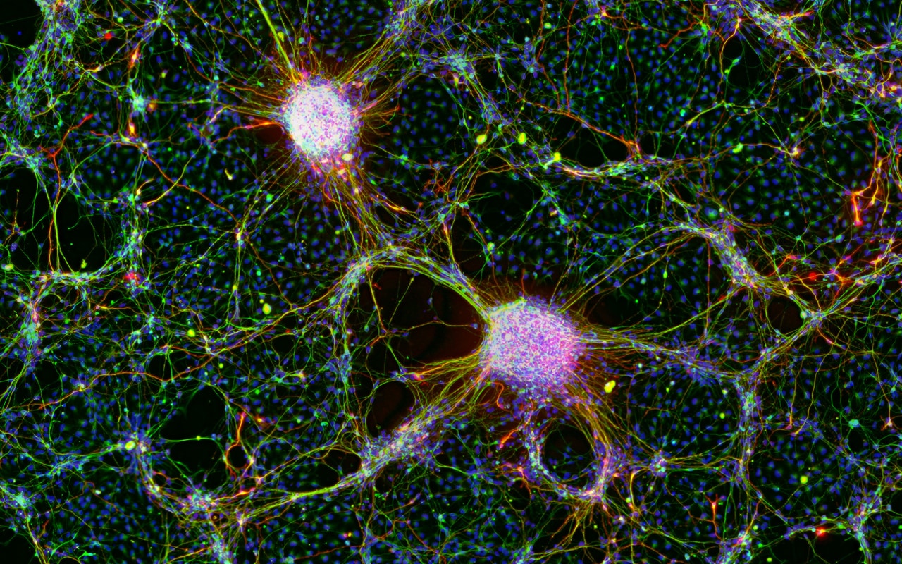
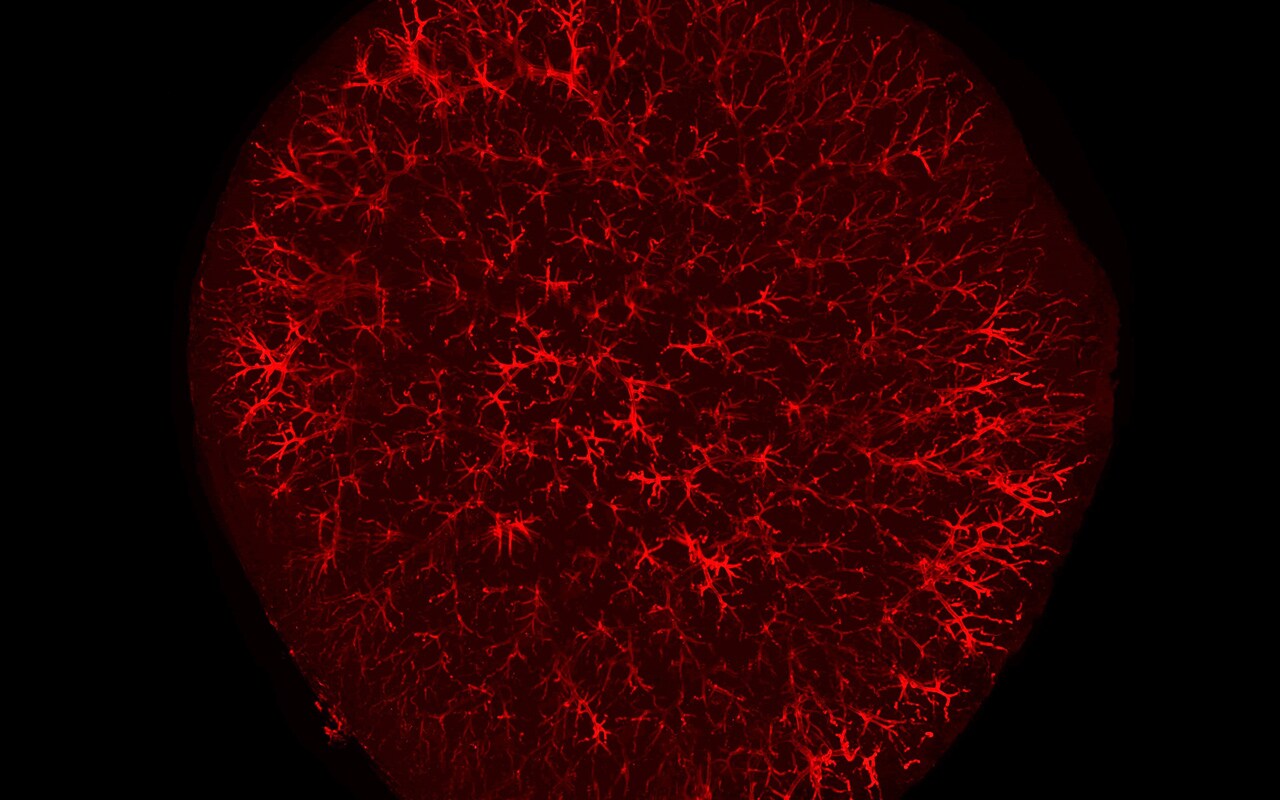
Human iPS cell-derived nerve cells
Courtesy of Assistant Professor Asuka Morizane, Department of Biological Repair, Field of Clinical Application, Center for iPS Cell Research and Application, Kyoto University
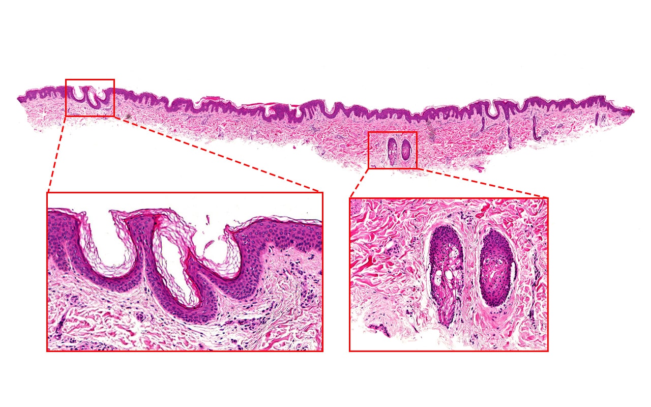
Understanding the Science Behind Fluorescence Microscopy
A fluorescence microscope functions in a different manner from a traditional optical microscope. This is because it utilizes fluorescence, which is the natural emission of light by fluorophores contained in a substance that has absorbed light or energy from another form of electromagnetic radiation. This can be a replacement for or in addition to the standard use of reflected or scattered light to create an image. By focusing light of a very specific wavelength onto a substance, the fluorophores emit light of a longer wavelength, creating the resulting image. Typically, samples are prepared by dyeing them with a fluorescent stain or the expression of a fluorescent protein.
Common applications for a fluorescent light microscope include research and quality control in the life sciences, medical, and biology sectors. The sensitivity and reliability of these instruments means they are used frequently to observe the presence of molecules inside cells as well as the location of individual cells within tissue. The fluorescence imaging systems developed by KEYENCE utilize light ranging from ultraviolet to near-infrared to analyze biological specimens or visually enhance 3D features at an extremely small scale. The BZ-X is also capable of optical sectioning microscopy, minimizing fluorescence blurring and providing clear optical slices of thick samples. A wide range of samples, including animal cells, plant cells, and cultured tissue can be easily observed.